Body

MRI use is key in diagnosing stroke

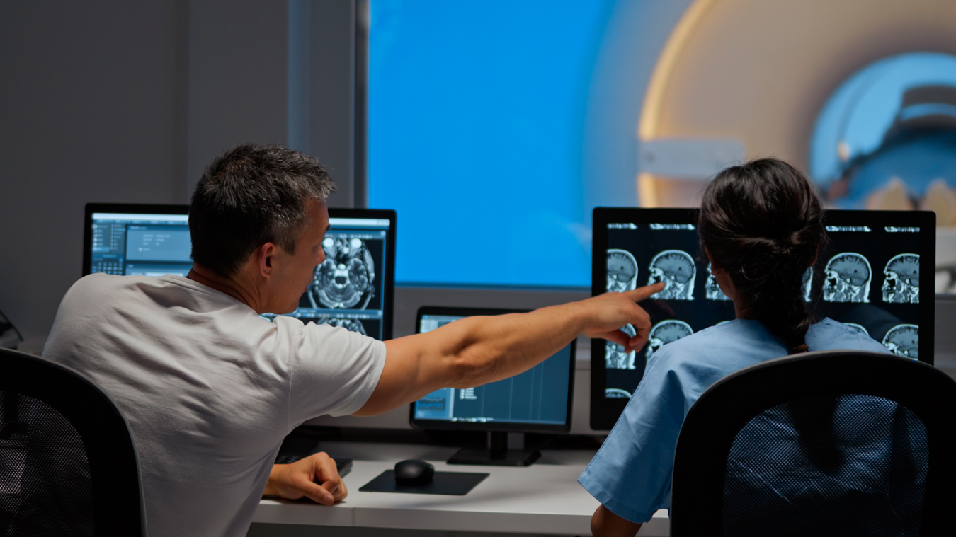
If a patient is suspected of having a stroke, their doctor may request a radiology test called magnetic resonance imaging (MRI). An MRI is an imaging procedure that uses magnets and radio frequency waves to take detailed pictures from deep within the body. It can target issues in bodily organs and tissues that otherwise appear healthy, like a blocked artery. An MRI provides crucial information to help make a stroke diagnosis, evaluate the best treatment options and predict the patient’s prognosis for the future.

An MRI is painless, noninvasive, requires no sedation or advance preparation and usually takes less than an hour. The MRI machine resembles a long cylinder that is open on both ends. Patients lie on a movable bed that goes into the scanner. Patients will be given headphones to block the loud clanking or tapping noises the scanner will make, and can ask for mild sedatives if they’re claustrophobic. 

How doctors diagnose strokes

A stroke occurs when the brain is deprived of oxygen. Strokes are typically classified as ischemic (when an artery in the brain becomes blocked) or hemorrhagic (when a blood vessel in the brain bursts, and the blood that’s released damages the surrounding brain cells). It’s important to quickly determine which type of stroke has occurred, because treatment options are different. The first test is usually a CT scan, which uses X-rays to show affected areas of the brain. A CT scan is effective for identifying the bleeding of a hemorrhagic stroke, but an MRI’s detailed images may be needed to spot the blocked area causing an ischemic stroke.

St. Charles Bend offers our patients a variety of advanced stroke treatments in Central Oregon.