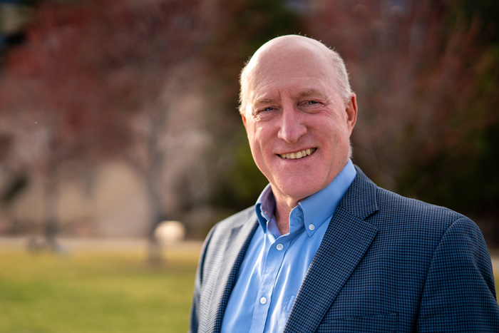
categories:
Body

St. Charles is increasing access for gastroenterology services in Central Oregon, thanks to a new partnership with WovenX Health, a virtual GI provider. 
 
Since the service began at the end of April, more than 200 patients have received care through it. Jonnie Becker, administrative director for the surgical division, explains that WovenX Health has provided tremendous relief for the gastroenterology department, which is routinely booked for months out. 
 
Patients must be referred to use the service. When they call to schedule an appointment with the GI department, patients may be offered on-demand appointments through WovenX Health, based on health-related criteria. Rather than waiting months, patients can connect virtually with a GI provider on the same day. Becker says it is also reducing patients seeking emergency care, as 15% of the patients using WovenX Health report they would have sought care in an Emergency Department if not for the on-demand visit availability. 
 
Becker says the process is seamless for St. Charles patients, and the response has been overwhelmingly positive. WovenX Health virtual providers are credentialed with St. Charles and are able to order supporting labs, imaging and procedures.  

Becker expects WovenX Health providers to see about 40 patients a week, which will also free up St. Charles providers to focus on procedures that cannot be done virtually, like scopes. 
 
“I am so pleased we have been able to expand our department with the collaboration with WovenX. This partnership has enabled us to increase capacity and offer GI services to many more patients,” said Becker. 

Share
topics in this article
Body

The spine is made up of small bones called vertebrae, which are stacked on top of one another. The bones connect to create a tube that protects the spinal cord. Spondylolysis is a common cause of low back pain in adolescents that is caused by a crack or stress fracture in one of the vertebrae. In some cases, stress fractures weaken the bone so much that it begins to shift or slip out of its place in the spine. This is known as spondylolisthesis.

Causes and risk factors

In children and adolescents, spondylolisthesis commonly occurs during periods of rapid growth (growth spurt). Spondylolisthesis is more likely to occur in young athletes who participate in sports that require frequent hyperextension of the lower spine, such as gymnastics, football, or weightlifting.

Symptoms

Common signs and symptoms of a vertebrae slippage include:

  • Back pain similar to muscle strain
  • Pain that radiates to the buttocks or back of the thighs
  • Pain worsens with activity and improves with rest
  • Back stiffness
  • Unusually tight hamstrings
  • Difficulty standing and walking
  • Tingling, numbness, or weakness in the legs

Treatment

Patients with a mild spondylolisthesis will usually improve with conservative treatment. Nonsurgical treatment options include:

  • Rest from sports or other activities that place stress on the lower back
  • Anti-inflammatory medications to help reduce swelling and relieve pain
  • Physical therapy exercises to improve flexibility and strengthen muscles
  • Bracing to limit the movement of the spine and allow fractures to heal

More severe cases of spondylolisthesis or cases that are not improving with conservative treatment may require surgery. A spinal fusion is the most common surgical procedure used to treat spondylolisthesis. Download and Print Educational Information About Spondylolisthesis

Body

Spina Bifida is a birth defect that occurs during fetal development when the spine does not form normally. As a result, the spinal cord, vertebrae, nerves and surrounding tissues may also be affected. The term spina bifida means “split spine” and it falls under the category of neural tube defects. The neural tube is an embryonic structure that will develop into a baby’s brain, spinal cord, and the tissues that enclose them.

Causes and risk factors

Normally, the neural tube forms early in pregnancy and closes by the end of the first month of pregnancy. This is when the spina bifida defect usually occurs. The cause of spina bifida is unclear, but it may be linked to a combination of genetic and environmental risk factors, such as:

  • A family history of neural tube defects.
  • A folate (vitamin B-9) deficiency
  • Diabetes and/or obesity

Symptoms

Spina bifida can occur in different forms. The severity of symptoms can range from mild to severe, depending on the type, size, location, and complications. 

Spina Bifida Occulta – “Occulta” means “hidden” in Latin, and this type of spina bifida is often called “hidden spina bifida.” The mildest form, spina bifida occulta results in a small separation or gap in one or more of the bones of the spine (vertebrae). Many people do not even know they have spina bifida occulta, though some may have pain or other neurological symptoms. A tethered cord, where the spinal cord is attached to surrounding tissue instead of floating freely, is a common complication of spina bifida occulta that may require surgery. 

Meningocele – In meningocele, the protective membranes that surround the spinal cord (meninges) push through openings in the vertebrae, forming a sac filled with nerve fluid (meningocele). The sac may be visible on a child’s head, neck, or back, and can happen anywhere along the spinal column or at the base of the skull. Because meningocele does not typically include the spinal cord, nerve damage is less likely. However, children with meningocele may have health complications if the nerves surrounding the spine are damaged. The severity of symptoms depends on the size of the meningocele and where it is located on the spinal column. 

Myelomeningocele or Spina Bifida Cystica – Known as open spina bifida, myelomeningocele is the most common and most severe form of spina bifida. It occurs when some of the bones in the back do not develop properly and do not close completely around the spinal cord. As a result, portions of the spinal cord, nerves, and surrounding tissue push through the incomplete backbone and are exposed. This often forms a sac that sticks out on a child’s back, typically exposing tissues and nerves. This can cause proneness to infection. Children also typically will lose nerve function below the location in which the defect occurs, leading to sensation loss, paralysis, and bladder problems. The paralysis can lead to deformities of the legs, feet, and back such as scoliosis. Children with myelomeningocele often have a buildup of cerebrospinal fluid in and around their brain because the fluid that protects the brain and spinal cord is unable to drain properly.

Treatment

Treatment for spina bifida depends on the severity of the deformity. Children with spina bifida occulta may not need any treatment, while children with myelomeningocele typically require surgery 1-2 days after birth to protect the exposed areas. Because spina bifida can involve many different body systems, your child may need support from a team of medical professionals.

Body

A superior labrum anterior to posterior (SLAP) lesion tear is an injury to the labrum of the shoulder, which is the ring of cartilage that surrounds the socket of the shoulder joint that helps stabilize it. A SLAP lesion tear occurs both in front (anterior) and back (posterior) of this attachment point. The biceps tendon can be involved in the injury, as well.

Causes and risk factors

  • Falling on outstretched arm
  • Forceful pull
  • Motor vehicle accident
  • Overuse
  • Repetitive overhead sports such as weightlifters or baseball pitchers
  • Throwing or trying to catch a heavy object
  • Shoulder dislocation
  • Aging

Symptoms

Symptoms of this injury can resemble many other shoulder issues:

  • Sensation of “catching”
  • Instability
  • Weakness
  • Limited range of motion
  • Pain when throwing or lifting objects

Treatment

Conservative treatments such as rest, ice, non-steroidal anti-inflammatory (NSAID) medication, and physical therapy can be effective. If these non-surgical treatment options do not improve pain, surgical treatment may be required. Commonly, this injury is repaired with arthroscopic surgery, which is when your orthopedic surgeon will insert a tiny camera and surgical instruments into your shoulder to repair it.

Body

The shoulder joint is described as a ball and socket joint, and is the most movable joint in the body. This allows the shoulder to provide the wide range of motion required to perform many activities, such as throwing, rotating, and reaching over our head. When the ball slips out of the socket, your shoulder has dislocated.

Causes and risk factors

  • Falling
  • Motor vehicle accident
  • Playing sports such as football, basketball, or volleyball
  • Forceful throwing, lifting, or hitting

Symptoms

A dislocated shoulder causes pain, and often an inability to move the shoulder. Other symptoms may include:

  • Instability and weakness
  • Swelling
  • Bruising
  • Visible bump under skin where ball has shifted
  • Numbness and tingling

Treatment

An orthopedic surgeon will reset the joint by moving your arm back into place. Your doctor may recommend rest, ice, a sling, rehabilitation exercises, and anti-inflammatory medications. Surgery is rarely needed for a first time dislocation, but it is often required for a shoulder that dislocates repeatedly (shoulder instability).

Body

Reverse total shoulder replacement is recommended for people who do not have a functioning rotator cuff. For these individuals, an anatomical total shoulder replacement can still leave them with pain and the inability to lift one’s arm past 90 degrees.

The design of this surgery is very innovative. As the name implies, the shoulder prosthesis does not recreate normal anatomy, instead the socket and metal ball are switched. That means a metal ball is attached to the shoulder bone and a plastic socket is attached to the upper arm bone. Patients receiving a reverse total shoulder replacement have a functioning deltoid muscle, which is the large muscle on the side of the shoulder, and a non-functioning rotator cuff, usually due to a large irreparable rotator cuff tendon tear. This procedure greatly improves the function of the shoulder by absorbing the some of the role of the absent rotator cuff. The prosthesis provides a stable and constant center of rotation, allowing the deltoid muscle to elevate the arm.

While this procedure does not make your shoulder normal, it does improve the function and reduces or eliminates shoulder pain. The prosthesis is non-atomic, but patients can expect to use the shoulder for light activities such as hunting, fishing, playing golf, riding horses, and doing lightweight training exercises in the gym. Most patients are able to drive within three weeks of surgery.

Body

What is a quadriceps tendon tear?

Tendons are strong cords of fibrous tissue that attach muscles to bones. The quadriceps tendon works with the muscles in the front of your thigh to straighten your leg.

Quadriceps tendon tears can be either partial or complete. Small tears of the tendon can make it difficult to walk and participate in other daily activities. A large tear of the quadriceps tendon is a disabling injury. It usually requires surgery and physical therapy to regain full knee function.

Causes and risk factors

Quadriceps tendon tears are not common. They most often occur among middle-aged people who play running or jumping sports. A quadriceps tear often occurs when there is a heavy load on the leg with the foot planted and the knee partially bent. Think of an awkward landing from a jump while playing basketball. The force of the landing is too much for the tendon and it tears. Tears can also be caused by falls, direct force to the front of the knee, and lacerations (cuts).

Symptoms

When a quadriceps tendon tears, there is often a tearing or popping sensation. Pain and swelling typically follow, and you may not be able to straighten your knee. Additional symptoms include:

  • An indentation at the top of your kneecap where the tendon tore
  • Bruising
  • Tenderness
  • Cramping
  • Your kneecap may sag or droop because the tendon is torn
  • Difficulty walking due to the knee buckling or giving way

Diagnosis and treatment

Your doctor will consider several things when planning your treatment, including:

  • The type and size of your tear
  • Your activity level
  • Your age

Most small, partial tears respond well to nonsurgical treatment including immobilization and physical therapy.

Most people with complete tears will require surgery to repair the torn tendon. Surgical repair reattaches the torn tendon to the top of the kneecap. People who require surgery do better if the repair is performed soon after the injury. Early repair may prevent the tendon from scarring and tightening into a shortened position.

Body

The posterior cruciate ligament (PCL) is a band of tissue that connects the femur to the tibia at the back of the knee joint. An injury to this stabilizing ligament requires a powerful force, like a motor vehicle accident or sports injury.

Symptoms

  • Knee pain
  • Instability
  • Swelling, stiffness, or bruising

Diagnosis and treatment

In many cases, a torn PCL will heal without surgery. Your orthopedic surgeon may recommend using the RICE method (Rest, Ice, Compression, Elevation), bracing, physical therapy, and taking non-steroidal anti-inflammatory drugs (NSAIDs). A doctor may recommend surgery if you have multiple injuries to your knee. Arthroscopic surgery is often used to rebuild the ligament. A torn PCL will be replaced with a tissue graft from another part of your body or a cadaver. Arthroscopic surgery is less invasive and results in quicker recovery times.

Body

The hip is a ball-and-socket joint. The rounded head of the upper femur (thighbone) fits firmly into the socket of the pelvis, and together they make up the hip joint. In newborns and children with developmental hip dysplasia (DDH), the hip joint has not formed normally and the ball of the femur is loose in the socket. This usually means the hip is very easily dislocated.

Causes and risk factors

DDH tends to run in families, and is more common in:

  • The left hip
  • Girls
  • Firstborn children
  • Babies born in the breech position
  • Family history of DDH
  • Oligohydramnios patients (low levels of amniotic fluid)

Symptoms

DDH is most often present at birth, though it may develop during the first year of childhood. Some babies born with DDH may not present any symptoms initially. Common signs of DDH include:

  • Legs are different lengths
  • Uneven skin folds on the thigh
  • Less mobility or flexibility on one side
  • Limping, walking on the toes, or waddling gait

Treatment

When DDH is diagnosed at birth, it can usually be treated conservatively with good results. If the condition is not diagnosed until a child begins walking, treatment can be a little more complicated. Nonsurgical treatment options include:

  • Harnesses to hold the hip in the proper position while allowing free movement of the legs
  • Bracing or casting are used if a firmer material is needed to keep the joint in the proper position

In some cases, the pediatric orthopedic surgeon will need to reposition your child’s femur into the right position, and apply a cast to hold the bones in place. This procedure is performed while the child is under anesthesia. Surgery to correct DDH is sometimes necessary to realign the hip. Download and Print Developmental Hip Dysplasia Educational Information

Body

However hard we try, it is impossible to keep our children out of harm’s way at all times. According to the American Academy of Pediatrics, fractures are the fourth most common injury in children under the age of six. Falls cause most of the reported fractures in children, but motor vehicle crashes are also a top contributor. A broken bone sounds scary, but pediatric fractures often heal without the need for surgical repair. Children’s bones are different than adults – they are softer, more flexible, and tend to absorb shock better. A child’s bone also heals much faster than an adult’s bone. Because their bones are still growing and heal so quickly, it is important to treat a fracture quickly and properly to avoid future problems.

Symptoms

  • Child may not report pain right away, but tend not to use fractured body part
  • Tenderness, numbness, or tingling
  • Trouble moving the injured body part
  • Swelling
  • Bruising or redness

If there are any physical deformities such as a bump or change in shape of the bone, or if the bone is visibly sticking out, it’s best to seek medical attention right away. Otherwise, it’s probably okay to wait to be evaluated until the next day. The St. Charles Center for Orthopedics & Neurosurgery NOWcare walk-in injury clinic provides fracture care and casting for all ages with no appointment necessary.